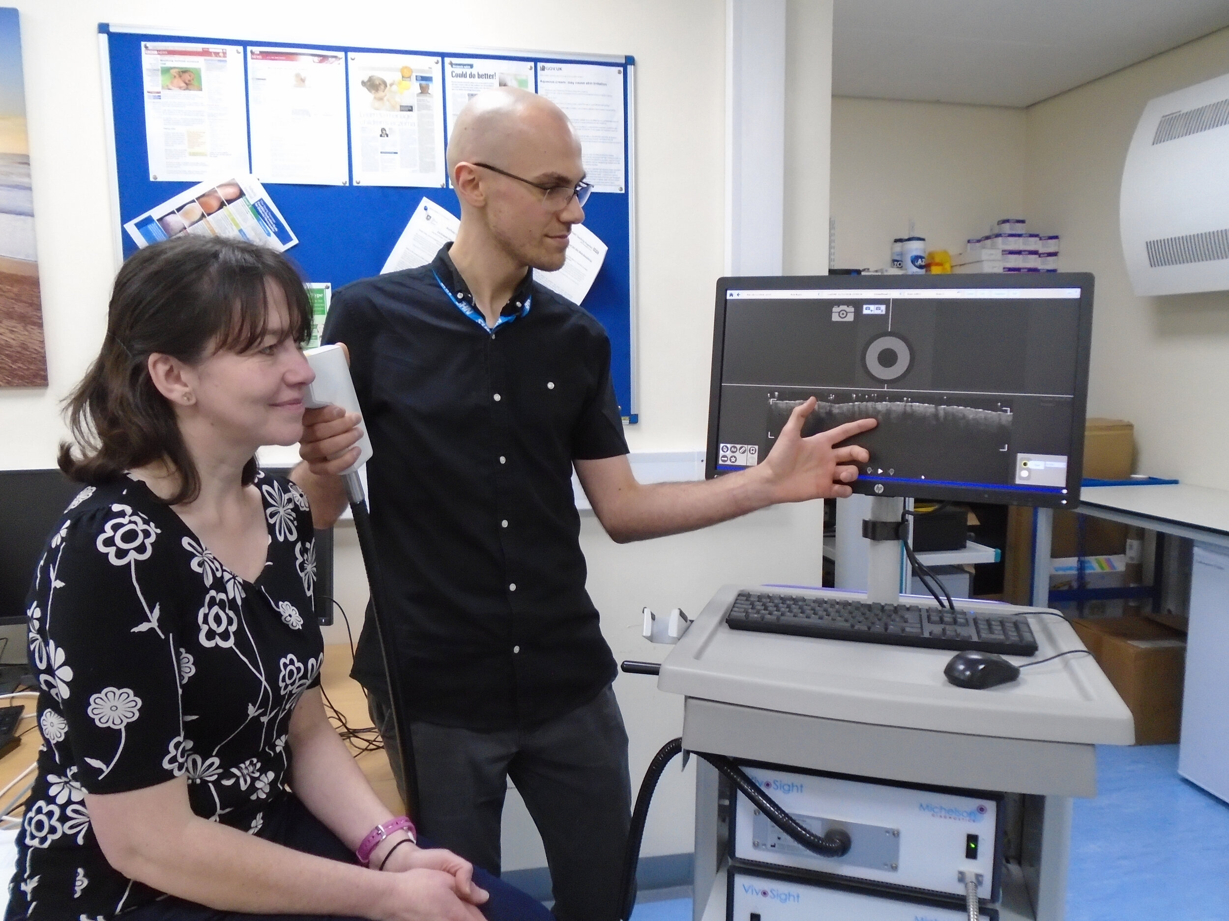
Optical Coherence Tomography (OCT)
Sheffield Dermatology Research conducts cutting edge OCT research within skin. Using modern optical instruments that shine light through the surface, we can see a detailed 3D map of sub-surface layers, vasculature and tissue structure.
What is OCT?
OCT is a non-invasive imaging tool, capable of visualising beneath the skin surface to a depth of up to 2mm. It is capable of imaging the epidermis and upper papillary dermis in micron scale resolution, capturing a 3D “optical-biopsy” of the skin sub-surface.
How does it work?
Light is beamed into the skin, the precise timing of returning reflections are then measured and used to build up a map of the internal tissue structure. This beam is scanned laterally in order to form a three-dimensional volume scan. The resulting data can be further processed in order to measure properties of the skin such as the location of blood vessels.
OCT for Atopic Dermatitis
The unique properties of OCT make it an ideal candidate for monitoring sub-clinical inflammation caused by atopic dermatitis (atopic eczema).
Sub-clinical inflammation occurs beneath the surface of the skin, is invisible to the naked eye and is ignored by traditional clinical grading systems. We need to be able to detect and quantify sub-clinical inflammation to optimise pro-active treatment regimens, induce remission and avoid the damaging and fluctuant course of atopic dermatitis.
Measuring sub-clinical inflammation
We currently use three unique types of OCT imaging to characterise aspects of sub-clinical inflammation.
Structural OCT
Provides a depth-resolved map of the tissue structure which can be used to study the skin layers.
Several key metrics of sub-clinical inflammation can be directly measured with structural OCT:
Mean Epidermal Thickness - Skin hypertrophy and thickening is associated with an increase in disease severity.
Skin Layer Area Roughness - A rougher stratum corneum is generally associated with increased skin dryness. A rougher dermal-epidermal junction is associated with the extension of dermal papillae in response to skin hypertrophy.
Skin attenuation coefficient - The rate of OCT signal decay with tissue depth is altered by the local tissue scattering and absorption properties.
In addition to skin thickening, skin thinning (Atrophy) is a key metric which can be used to evaluate the safety profile new topic therapies against that of topical corticosteroids.
Over a 4-week, twice daily treatment period, Betamethasone Valerate (0.1%) cream (BMV), a potent topical corticosteroid, demonstrated significantly higher epidermal atrophy than Tacrolimus (0.1%) ointment (TAC), a topical calcineurin inhibitor.
Angiographic OCT
Captures the morphology of microvasculature within tissue by analysing the structural signal transiently.
The vasculature within skin forms a complex three-dimensional structure consisting of rising capillary loops being fed by a dense horizontally arranged superficial plexus.
Alterations in blood vessel morphology and depth occur in tandem with skin hypertrophy. As the vessels are only present within the dermis, their most superficial location can be used as a lower-bound of the dermal-epidermal junction location. In cases of poor structural OCT contrast, where discerning this layer can be challenging, this represents a potentially more robust, surrogate marker of epidermal hypertrophy and thickening.
Several key metrics of sub-clinical inflammation have been identified using angiographic OCT:
Superficial Plexus Depth - The depth of the horizontal superficial plexus within skin. Measured as the point at which capillary loops begin to interconnect between each other. This layer is pushed downwards as the epidermis thickens.
Capillary Loop Depth - The upper depth of capillary loops within the skin. This appears to be subtly increased with skin inflammation, despite the extension of dermal papillae.
Mean Vessel Diameter - The average thickness of vessels within the superficial plexus, which appears to increase with inflammation.
Vessel Fractal Dimension - The tortuosity, or “twistedness” of vessels in the superficial plexus which appears to increase with disease severity.
Polarisation-sensitive OCT
Provides additional contrast in skin by measuring the time delay between returning light consisting of two orthogonally polarised states. Light travelling through highly aligned fibres (Such as collagen) accumulates time delay, or phase-retardance, between these polarized states, leading to a visible banding pattern with polarisation-sensitive OCT, a phenomenon known as birefringence. Comparatively light moving through regions of disorganised, or damaged fibres have no such banding.
The animation above shows a central region of intense phase-retardance banding, which is invisible to structural OCT. In this example, the banding is caused by a striae alba (skin stretch mark) which has resulted in the alignment of collagen fibres and an increase in localised birefringence.
Skin Collagen Index - A metric which quantifies the degree of phase-retardance banding for a particular location. With atopic dermatitis we expect to see a reduction in the local Skin Collagen Index when compared to healthy skin, as a result of the local tissue damage and fibre disorganisation that is characteristic of inflammatory skin conditions.
In summary
OCT is a powerful imaging tool which facilitates a deeper look at the subclinical state of the skin. It provides a unique balance between resolution and depth penetration that is ideal for the study of subsurface skin layers and microcirculation.
In addition to atopic dermatitis, there are a wide range of potential applications for OCT in dermatology:


